
Search
Search Criteria
Products meeting the search criteria

HyPort 9000
With the growing needs of healthcare and the constant focus on improving workflows, the most demanding customers of modern technology are always patients and healthcare facilities.
Mindray met their needs by developing the HyPort range of medical supply units that combines cutting-edge technology with ergonomic use.
Surface protection by anodic oxidation / powder coating.
Rail for quick connection of connections.
Two brake systems for maximum safety.
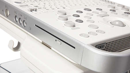
Ergonomic touch control panel.
Multi-screen information system.
A special tripod for attaching the anesthesia machine.

18 studies report excellent TRIOS accuracy…
18 independent studies report statistically higher accuracy for TRIOS compared with conventional impressions and/or impressions made with other major intraoral scanners for full arch, single unit and multi-unit restorations) 18 in vivo and in vitro studies (between 2015 and 2018).
Award winning technology…
3Shape TRIOS intraoral scanner has received Cellerant “Best of Class” 2018 Award for intraoral scanners. This marks a record-breaking six years in a row that TRIOS wins the “Best of Class” award in the digital impression solution category.
Here are the benefits at a glance...
- Caries Diagnostic aid built in - NEW for TRIOS 4 - Detect surface caries with fluorescent scans and interproximal caries with infrared scan - with no radiation.
- Smart tips - be scan ready in seconds - NEW for TRIOS 4 - Instant heat smart tips so you are always scan ready in less than 40 seconds, giving an additional 30% battery life and ability to scan 2/3 times as many patients per battery.
- TRIOS patient monitoring for preventive insights – NEW for TRIOS 4 - Scan every patient every time to accurately track changes in teeth and identify dental conditions sooner. Share visual monitoring information with patients to advance their understanding of issues and the need for treatment.
- AI scan technology for simplified scanning - Removes unnecessary soft tissue as you scan to make scanning easier.
Surface Caries
TRIOS 4 has built-in fluorescent technology that aids in the identification of possible caries. Using TRIOS 4, dental professionals can now be aided in the early-detection of surface caries, without the need for an additional scanning device.
Interproximal Caries
TRIOS 4 will also feature a dedicated transillumination smart tip later this year. The smart tip will aid in the identification of possible interproximal caries undetectable to eye, without emitting radiation, and is the first launch within 3Shape’s new smart tip platform.
- Wireless innovation for enhanced comfort and ease - The world’s first wireless scanner option enables you to scan unrestricted by wires, optimizing comfort for both you and your patients, and making scanning easier.
- Realistic colors and shade measurement for patient engagement - Create high-quality digital impressions in lifelike colors and apply shade measurement to evaluate treatment and activate quality dialogue with patients.
- TRIOS Patient Specific Motion to perfect your restorations - Record a series of different bite positions and highlight occlusal contacts for greater insights into case possibilities.

EP-1000 is an ophthalmologic diagnostic unit for a complete examination of the retina’s function, the visual pathway, and the optic nerve. A variety of diagnostic procedures can be performed, including ERG, VEP, MERG, MVEP, and EOG. This unit can only be operated by physicians and medically trained technical assistants. EP-1000 complies with the ISCEV’s standards (International Society for Clinical Electrophysiology of Vision).
Computerized Electrophysiology Device Tomey EP-1000 Multifocal, NEW!
Whenever you need detailed information,
the Multifocal will be your first choice
The EP-1000 Multifocal (mf) allows you to do all mf standard tests:
mfERG flash (FOK, SOK, flicker)
mfERG pattern (9 pattern for stimulation)
mfVEP (dartboard stimulation) for analysing local retina functions
Accessories and Optionals Analyses can be displayed in curves, values, 2D, 3D, rings, quadrants, groups and table.
Cross-target for macula dystrophy patients time
Mf pattern ERG with scrolling text target
Full access to all settings
e. g. position of the fixation target
No matter if you want to receive standardised clinical data or are heading towards a more scientific
oriented environment Tomey offers you the perfect individual electrophysiology solution.
The EP-1000 Multifocal is based on short M-sequences. This allows you to re-check all conditions of the exam, such as proper fit of electrodes or acceptable responses within a very short time (8 sec.).
With the fixation control hexagon the results can be controlled after every cycle. You dont have to wait until the end of the whole examination for controlling the fixation of the patient.
Different fixation targets are available: the cross-target covers the whole stimulation monitor to make exams possible for macula dystrophy patients. The changing
animal target is for children and the scrolling text target for all other patients.

Digital Fundus Camera Canon CF-1 inkl. Canon EOS-50D Mark II MED, NEW!
Overview:
The CF-1 Digital Mydriatic Retinal Camera is easy to use and efficient for both doctor and photographer. When used with a Canon EOS® digital SLR camera (sold separately), users can enjoy renowned Canon optical technology for superb imaging quality. User-friendly control software helps to ensure efficient workflow, potentially resulting in increased patient throughput. The CF-1 Digital Mydriatic Retinal Camera provides high-resolution color, red free, and fluorescein angiography imaging performance and heightened efficiency for eyecare professionals.
Features
Color, Red-Free, and Fluorescein Angiography Imaging
The CF-1 Digital Mydriatic Retinal Camera integrates a digital SLR camera from Canon's EOS series to achieve high resolution images with excellent detail, contrast, and color fidelity.
Easy Alignment and Focus
The 50-degree view angle enables easy alignment and focus with less flash flare, even with smaller pupil sizes. Only a low amount of light is needed to capture clear images.
2x Digital Magnification
Zoom the retina to 2x magnification and the system automatically crops out the peripheral edges of the image so the region of interest is larger in the frame.
Ergonomic Control Panel
Operator controls for key features such as shutter release, lamp setting, ISO adjustment, and mode switching are grouped together for one-handed operations in darkened exam rooms.

Premium technologies bring a new level of image quality to compact ultrasound so performance isn't sacrificed for portability. Designed for critical study requirements and big system performance everywhere you need it.
Wireless & wired DICOM for connectivity in any environment
PureWave Everywhere
Expand diagnostic information
Portable ultrasound when fast action is needed
Digital broadband beamforming on a compact
On cart
SonoCT brings a new level of image quality
SmartExam reduces exam time by up to 50%*
Remote travel
XRES brings a new level of clarity to compact ultrasound
Fine‐tune exams with active native data
Premium performance in the surgery suite

Mindray puts its latest technology in the Mindray DC-70 ultrasound device. This technology called X-Insight provides detailed clinical information about customers and is combined with advanced ultrasound technology.
The Mindray DC-70 is designed to operate at high efficiency with precise image accuracy enabled by eXpress Clarity and eXceptional Intelligence technologies and the benefits of the eXceeding Experience.
eXpress Clarity
Better Image Clarity
X-Engine
The latest X-Engine technology integrates a GPU and CPU capable of parallel processing for fast images. With state-of-the-art drawing engines, processing speeds are up to three or four times faster than traditional methods.
New iLive with Hyaline
This upgrade from iLive technology increases the resolution and authenticity of detailed anatomy. Hyaline is a new rendering method that provides dynamic transparency of the render structure to display a more comprehensive anatomical image so that the internal anatomy is clearer.
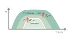
Single Crystal Probe Transducer with 3T . Technology
Combining Mindray's unique 3T technology (Triple-matching layers, Total-cut design, Thermal control), the convex and phased array probes are able to provide wider bandwidth as well as deeper penetration and higher resolution resulting in an optimum scanning solution in OB/ GYN, ABD, Cardiology and more.
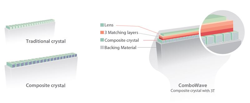
ComboWave Transducers
Compared to traditional probes, the ComboWave probe utilizes a piezoelectric composite material to optimize spectrum and reduce acoustic resistance. Integrated with Mindray's unique 3T technology, the ComboWave linear transducer probe enables you to achieve incredible performance with extreme image resolution in lymph nodes, chest, vasculature and more.
eXceptional Intelligence
Smart Planes CNS
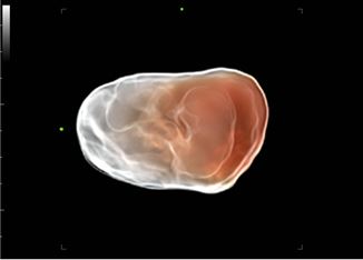
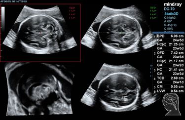
Smart Planes CNS is a user-friendly tool that improves scanning accuracy and fully automated operation thereby providing accurate diagnosis, better results and less operator dependence. With a simple button on the 3D image of the fetal brain volume, standard CNS results (MSP, TCP, TTP, TVP) and associated anatomical measures (BPD, HC, OFD, TCD, CM, LVW) will be obtained immediately with high accuracy.
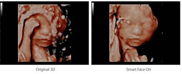
Smart Face
Smart Face provides intelligent and fast optimization of the fetal face with just one button. This feature can remove obstructions such as the umbilical cord, placenta, uterus so as to produce an optimal view of the fetal face.

Smart FLC
Automatically detects number and counts bags from 3D volume images.
- Accurate bag size assessment
- The bags are automatically sorted by size by color coding
- Easy reports with color graphics for studying pockets
Smart OB
Automatic fetal parameter measurement: trace and calculate BPD, OFD, HC, AC and FL with one click.
Smart NT
Automatic tracing of tube cavities with measurement results.
eXceeding Experience
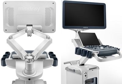
Eagle-Wing Arm with 21.5" / 23.8" Full HD Monitor
The exclusive Mindray eagle-wing provides unlimited settings for flexible monitor positions according to clinical needs.
13.3" Slim multi-gesture touchscreen with adjustable viewing angle
Gesture operation brings the latest trend in cart-based ultrasound devices with an intuitive and intelligent user experience beyond your expectations.
Transducer Probe Options
Convex (Convex / Convex)


Micro-Convex

Volume 4D Probe


Endocavity / Transvaginal


Bi-Plane


Pencil / Pedoff

linear


Phased Array


Corneal biomechanical properties influence the results and outcomes of ocular measurements and procedures, and may hold clues to diagnosing and managing ocular diseases.
Until now, assessing the biomechanical properties of corneal tissue has not been possible, confining practitioners and researchers to measuring purely geometrical aspects of the cornea, such as thickness and topography.
The Reichert Ocular Response Analyzer provides a new measurement of corneal tissue properties called Corneal Hysteresis (CH) that is a result of viscous damping in the corneal tissue.
How does it work?
The Ocular Response Analyzer utilizes a rapid air impulse, and an advanced electro-optical system to record two applanation pressure measurements; one while the cornea is moving inward, and the other as the cornea returns. Due to its biomechanical properties, the cornea resists the dynamic air puff causing delays in the inward and outward applanation events, resulting in two different pressure values:
The average of these two pressure values provides a repeatable, Goldmann-correlated IOP measurement (IOPG). The difference between these two pressure values is Corneal Hysteresis (CH); a new measurement of corneal tissue properties that is a result of viscous damping in the corneal tissue. The ability to measure this effect is the key to understanding the biomechanical properties of the cornea.
The CH measurement also provides a basis for two additional new parameters: Corneal-Compensated Intraocular Pressure (IOPCC) and Corneal Resistance Factor (CRF). IOPCC is an Intraocular Pressure measurement that is less affected by corneal properties than other methods of tonometry, such as Goldmann (GAT). CRF appears to be an indicator of the overall resistance of the cornea.
Product Features:
Comprehensive Pressure and Cornea Information
*
IOPCC (Corneal Compensated IOP) - New IOP measurement less affected by corneal properties.
*
CH (Corneal Hysteresis) - Measure of viscous damping in the cornea, providing new indicators for disease diagnosis and management.
*
CRF (Corneal Resistance Factor) - Measure of the overall rigidity of the cornea.
*
IOPG (Goldmann Correlated IOP) - Traditional IOP measurement for historical reference.
Advanced Technology
*
Patented dynamic bi-directional applanation process enables the only direct measure of corneal biomechanical properties.
*
Eliminates operator variance by utilizing microprocessor control technology and objective measurement criteria.
*
Patient and Operator Friendly
*
No chinrest and a soft, quiet air puff make the Ocular Response Analyzer a comfortable, non-intimidating experience for your patients.
*
Fully automatic alignment and measurement provides fast, repeatable results, allowing any member of your staff to conduct an exam.
Powerful Software Package
*
Enables automatic data capture of all measurements for analyzing trends, treatment efficacy, and diurnal variations.
*
Comprehensive patient management database simplifies record keeping.
*
Review results for a single patient, or data for an entire group of patients for population statistical analysis.
*
Comprehensive printout for hard-copy results.
*
Export data for use in other applications.

bon COBRA 5. MP non-mydriatic digital fundus camera, NEW!
Incl. chinrest and software
COBRA - The next generation of non-mydriatic fundus camera.
Content:
Cobra fundus camera incl. chin rest
Phoenix suite software
b o n delivers the innovative non-mydriatic digital fundus camera that integrates every function required for easy retinal screening.
Integrating an innovative imaging optical system, Cobra realizes digital fundus imaging of high resolution and fine gradation.
Ergonomically designed Cobra provides clear and detailed display of the entire fundus image at true 60° x 45° field of view.
The system offers retinal photography with minimum flash exposure allowing quick and efficient fundus photography, thereby minimizing patient discomfort.
Cobra shares the use of the high resolution CCD camera for alignment (IR illumination) and capture (White light flash).
Non-mydriatic
Cobra only needs a minimum pupil diameter of 2,2 mm and a room with standard lighting conditions.
Anterior Segments
Thanks to its variable focus options Cobra also provides pictures of the anterior segments of the eye.
High-speed image transfer to PC
Firewire connection to a PC allows quick and easy transfer of the images. The data are saved in a database through the Phoenix Software in Stand-Alone or network configuration.
DICOM connection can be achieved transferring images to a compatible server.
Phoenix Software imaging and archiving features
Sophisticated imaging functions are incorporated, including image processing features, drawing, measurement, and panoramic imaging for large field analysis.
With Phoenix it is easy to print and save structured reports on the patient archive.
Image export is available in many image formats. The data can be transferred to a DICOM (Digital imaging and Communications in Medicine) compatible server.
Imaging features
# Zoom effects
# Color control and filtering
# Measurement
# C/D ratio, Disc HV, Cup HV
# Drawing (text / objects can be inserted)
# Edge enhancement
# Grey scale
# contrast RGB
# Red-free and channel splitting
# Color inversion
# Brightness, Contrast and Gamma control

Automated Perimeter Kowa AP-7000, NEW!
Automated perimeter for both static and kinetic perimetry
Next generation perimeter with the gold standard threshold test
The AP-7000 is Kowas next generation automated perimeter that offers an extensive variety of test strategies and screening
programmes. It provides reliable and consistent assessment results due to its normalised database.
Full threshold modes offer macular, central and peripheral coverage up to 80° whilst screening modes provide swift evaluation of the visual field for relative or absolute scotomas. To shorten test times quick modes are available for both threshold and screening modes. Furthermore, by automatically correlating the fundus image (from a fundus camera, OCT or SLO) with the static visual field, early detection of glaucoma is possible.
With its ergonomic, compact design, the AP-7000 offers additional comfort to patients, easy operation for the clinician and fits perfectly within the practice.
Fundus Oriented Perimetry
Static perimetry test can be applied to abnormal sites on a fundus images, such as a fundus photograph, OCT or SLO.
Threshold test within the central 10°, custom optional threshold that allows selection of any desired test point, and custom optional screening tests are available.
Threshold Macula 2 test, Custom opticonal threshold test and Custom optional screening test are available.
Threshold Macula 2 test examins within the central 10°, Custom optional tests allow selection of any desired test points.
In addition to test within the central 30° that can observe the progression of glaucoma, test is possible in a wide variety of range, including central 10°, which can identify visual field abnormalities in the macula.
Analytical Indices
GHT (Glaucoma Hemifield Test)
For this index, threshold center test points are divided into ten sectors, and corresponding sectors above and below the axis of the horizontal median are compared.
VFI (Visual Field Index)
A percentage index in which a normal visual field is 100% and total loss of field is 0%
Anderson's Criteria Diagnostic Support Function
If one or more of three consecutive points satisfy one or more of the conditions "PSD has p<5%", "GHT outside normal limits", or "patern deviation probability plot shows a cluster of three or more nonedge points that have p<5%, and one of the points has p<1%" this indicator judges the condition to be a glaucoma visual field abnormality. (The physician must judge whether the three points match the NFL movement.)
Chronological Change Display
Test result analytical indices can be graphically displayed as time series data to give a clear grasp of changes over time in the tested eyes.
Predictive Display
Predictive graphs are displayed from calculations of linear rates of changes in analytical indices. This function predicts what values of MD and VFI (Visual Field Index) will be reached at what age, if current rates of change in those values continue.
Convenient Tabbed User Interface
Main operation buttons are grouped at the top of the page, and buttons are laid out to follow the progression of tests, from patient information entry through test program selection to result display.
4-zones measurement that goes beyond screening, and programs using probability values (p-values) in intensity steps, are among the features that enable effective test in less time.
Running tests with intensity of probability value (p-value) makes it possible to display the difference between the measured value and the normal value for each age as a p-value, so that evaluation equivalent to total deviation in thresholds can be performed in a shorter time.
"Automatic measurement function", using many median patterns, "Manual measurement function", allowing free drawing of isopters, and "auto + manual measurement function", which allows any drawing of median after automatic measurement, are among the diverse measurement method options available.

The EM-4000 offers non-contact examination, auto-alignment and measurement, with automatic analysis of the endothelium layer. The EM-4000 is professional and quick (4 seconds for both eyes). The auto-alignment technology means reproducibility of the measured areas in insured. The integrated non-contact pachymetry automatically measures with every central examination. The large colour touch screen is used as an operating monitor as well as for displaying all measured values. The EM-4000 has a very large measurement area, with up to 300 counted cells the system assures a representative cell density analysis of your patients’ cornea. Images can be taken at 13 position (the centre and 12 peripheral points). The thickness of the cornea is automatically measured with every central exam. The software evaluates all relevant data respective to the endothelium, such as the density of the cells as well as Polymegathism and Pleomorphism (morphology). High quality images enable discovering irregularities or degeneration of the endothelium possible. For difficult cases you can use the classical L-count function, the Core method and Tomey’s special Dark Area analysis tools.
EM-4000 Features:
- Auto-alignment & auto-measurement
- Integrated non-contact Pachymetry
- 13 measurement areas
- Integrated database
- L-Count, Trace, Core and Dark Area methods
- Counts up to 300 cells
- Integrated printer

b o n PERSEUS Specular Microscope
Non-contact examination, auto-alignment and automatic vertices-based analysis of the endothelium layer within a few seconds, makes working with the Perseus smart, professional and accurate.
Friendly user interface: the examination is performed via very user-friendly touch screen alignment.
Wide measurement area: the wide measurement area makes it possible to count up to 300 cells (average value on normal eyes) and enables the analysis software to get a reliable cell density along with the analysis of cell size and shape.
Minimum light, maximum comfort: Perseus ensures maximum patient comfort due to the low intensity the light source.
Seven measurement areas: images can be taken at 7 positions (single centre position and six peripheral points).
Pachymetry: non-contact pachymetry is performed on every acquisition
High magnification during analysis makes it possible to zoom into the image seeing the limit of each cell in detail.
This enables the user, via touch screen navigation, to edit individual cells.
Software features: the intuitive software evaluates all relevant endothelium data such as
Number of counted cells
Cell density (number of cells per 1 mm2)
Average dimension of analysed cells
Standard deviation of analysed cell dimension
Coefficient of variation
Statistical distribution of sizes (polymegathism) and shapes (pleomorphism)

ZEISS modern vision testing at its best
Skillful determination of best-corrected visual acuity can be time-consuming, requiring operator experience and various fine adjustments. It is well worth the effort when the result is perfect vision correction, a superior viewing experience and highly satisfied patients – as with the digital phoropter VISUPHOR® 500 and the acuity chart systems VISUSCREEN 100 and VISUSCREEN 500 from ZEISS. Combining comfort and convenience with speed and efficiency, they offer refraction specialists a sophisticated, modular solution.
Made to assist your expertise
> Flexible: Subjective refraction systems from ZEISS are modular and expandable to suit your needs.
> Simple: Acuity charts and phoropter allow easy, intuitive operation via touch-screen interface.
> Efficient: Preconfigured workflows let you get right to work, saving you time preparing refraction tests.
> Connected: You can upload presets from the autorefractor VISUREF® 100 and the digital lensmeter VISULENS® 500 from ZEISS; or directly connect to selected data management systems such as FORUM® from ZEISS.
Simple touch screen control
Simplicity best describes the handling experience of the ZEISS subjective refraction system. The clearly structured user interface makes working with the ZEISS VISUSCREEN/VISUPHOR very easy. With the fully integrated GUI, both the phoropter and acuity screen can be controlled as one subjective refraction unit. Even single-handedly, with the optional, medical grade panel PC or an iPad.
Smart workflow
The ZEISS VISUPHOR 500 enables preconfigured workflows. Thereby, settings for each test are optimally adjusted, fully automatically, making standard refraction tests simple and fast. Settings and workflows can also be individually configured in the freestyle mode. Patient contact with the headrest is continuously monitored and displayed on the screen.
| Technical data | ||
|---|---|---|
| Spherical lenses | –19.00 to +16.75 D (increments: 0.12 / 0.25 D) | |
| Cylinder lenses | 0 to ± 8.75 D | |
| Cylinder axis | 0 to 180° (increments: 1° steps) | |
| PD | 48 to 80 mm | |
| Rotary prism | 0 to 20 ∆ | |
| Retinoscopy | +1.5 D, +2.0 D | |
| Pin hole lens | 2 mm | |
| Maddox rod | Right eye: red, horizontal / left eye: red, vertical | |
| Red/green filter | Right eye: red / left eye: green | |
| Polarizing filter | Right eye: 135°, 45° / left eye: 45°, 135° | |
| Split prism | Right eye: 6 ∆ BU Left eye: 10 ∆ BI (up to 5 ∆ complement) | |
| Line voltage | 100 – 120 / 200 – 240 V AC ± 10%, 50…60 Hz | |
| Power consumption | 145 VA | |
| Dimensions (W x H x D) | 361 x 280 x 108 mm | |
| Weight | 5 kg | |
| Interfaces | 2 x RS232 / Bluetooth | |
| System requirements | iPad or iPad Air with iOS 11 or better | |

Incl. Nikon SLR and VK-2 software
Features
KOWA nonmyd 7 FUNDUS CAMERA
Digital 16 Mega-Pixels External Integrated Camera Back
Nonmyd Color 45 degree / 20 degree Optical Magnification
Small Pupil Mode
Multiple Step Flash
Focusing/Alignment Dots
Easy to Operate
Specifications
Picture Angle 45 Degrees / 20 Degrees Optical
Working Distance 30mm (distance of examined eye to front end of objective lens)
Minimum Pupil Diameter 3.7mm
Focusing Split Luminous Bars
Working Distance Adjustment 2 Luminous Dot Indication Types (Anterior Chamber / Fundus Electrically Switched)
Internal Fixation Lamp 3 Positions (Central, Nasal, Temporal)
External Fixation Lamp Red Light (Option)
Monitor 5.5" LCD Monitor
Interface IEEE1394
Power-Saving Function Timer
Optical Head Movable 40mm Forward / Backward, Movable 100mm Leftward / Rightward, Movable 30mm Vertically (Electric)
Chinrest Moving Distance Movable 60mm (Electric)
Power Supply Input: AC100V - AC240V, 50 / 60Hz
Dimensions 310 (W) x 504 (D) x 548 (H) mm
Weight 21Kg / 46 Ibs.
VK-2 DIGITAL IMAGING SOFTWARE
Fast Patient Set-up
On Screen Stereo Viewing
Easy Operation
Multi-video Input
FA / Color / ICG / Slit Lamp
Complete Image Manipulation
Bridge Integration with OfficeMate and other popular electronic record management software
Network Ready
Telemedicine Ready
True Merge Function

Digital NM/M/FAG Retinal Camera Kowa VX-10α, NEW!
Two-In-One Mydriatic and Non-Mydriatic Fundus Camera
High functionality, high-quality images and easy to use, VX-10α is the ideal tool for eye-care photography.
VX-10α becomes the perfected fundus camera performing all non-mydriatic color, mydriatic-color and FA.
Please find all technical detail at the PDF belonging to this offer.

Scheimpflug Topographer Tomey TMS-5, NEW!
* High resolution
* Anterior + posterior map
* Pachymetry map
* Anterior chamber depth
* High speed measurement: 0.3 sec.
* Operates in all light conditions

bon "SIRIUS" - High Resolution 3D Rotating Scheimpflug Camera and topography system,
including software, electric table, computer and TFT display
The excellent combination between a rotating Scheimpflug camera and a Placido disk technique provides the best performances for measuring:
# Anterior and posterior corneal topography
(sagittal and tangential, curvature and elevation maps)
# Corneal Pachymetry (12 mm diameter)
# Anterior chamber analysis
# OPD analysis and visus simulation
# Keratoconus screening
Device features
# Refractive and cataract surgery planning tools
# Glaucoma screening
# Keratoconus screening
# IOP correction formulas
# Optical cornea analysis
# Cataract summary
# Glaucoma Summary
# Keratoconus Summary

Digital Hybrid Digital Fundus Camera Canon CX-1
Complete workstation incl. Digital Hybrid Digital Fundus Camera Canon CX-1 incl. Canon EOS-60D MarkII MED, Power-PC, HD-screen, electr. ophthalmic table, NEW!
The CX-1 is a MYD and Non-MYD hybrid digital retinal camera. It is extremely versatile with its 5 photography modes; colour. FAG, red free, cobalt and FAF (Fundus Auto Fluorescence). Making it ideal for screening and the diagnosis of the main eyes diseases.
Features
* Digital Retinal Camera,
* Hybrid camera MYD / Non MYD
* FAF (Fundus Auto Fluorescence), also in Non Myd
mode
* Use of EOS technology
* Advanced Stereo photography system
* Bundled Retinal Imaging Control Software
* Open connectivity and DICOM compliant
Canons CX-1 is a compact and portable hybrid retinal camera that combines both Mydriatic and Non-Mydriatic Modes, and switches between the two with a simple touch of a button.
Hybrid camera
Canons CX-1 is a compact and portable hybrid retinal camera that combines both Mydriatic and Non-Mydriatic Modes, As well as saving on equipment investment, this function also means that the examiners using the equipment can screen for more than one disease, e.g. AMD (Age-related Macular Degeneration), Glaucoma and diabetic retinopathy, with the same unit. This saves time and removes the need for repeat appointments.
5 Photography Modes
With the CX-1 following photography modes are available; Color, Red-free, Cobalt, Fluorescein Angiography and Fundus Autofluorescence (FAF) Alignment and observation with the CX-1 can be done through the viewfinder or by the large EOS LCD screen
FAF (Fundus Auto Fluorescence)
Detecting Auto fluorescence of the retina is an important indication of the retinas health. With the CX-1 it is possible to take FAF (Fundus Auto fluorescence) images, even in Non-Mydriatic mode.
Use of EOS Technology
Canons own EOS camera technology, with its renowned image processing capabilities, is adapted exclusively for medical use in the CX-1 to provide optimal retinal imaging in a compact and convenient system. The single onboard 15.1 MegaPixel digital camera handles five different photography modes with ease, including non-mydriatic FAF photography, allowing EOS imaging technology to benefit all retinal images from the CX-1.
Enhanced stereo photography function
Easy capture for stereo view, by using the stereo guide marks on EOS LCD in the non-mydriatic mode, or stereo unit (option) a stereo pair can be created and managed very simply
Bundled Retinal Imaging Control Software
Bundled Retinal Imaging Control Software (RICS) for full camera control and image optimalization. The software has extensive diagnostic tools for optimized workflow and patient management; e.g. Cup/Disc ratio calculation, compare studies, RGB channel display, stereo viewing
Open connectivity and DICOM compliant
The CX-1 with RICS has open connectivity with existing networks and is fully DICOM compliant.

Digital NM/M/FAG/ICG Retinal Camera Kowa VX-10i, NEW!
Two-In-One Non-Mydriatic and Mydriatic Fundus Camera.
Highly effective and ergonomically designed, the ideal tool for eye-care photography.
VX-10i, camera of extend possibilities with the ICG filter, VX-10i become the perfected fundus camera performing all non-mydriatic color, mydriatic color, FA and ICG.

Overview
The Canon CR-2 PLUS Digital Non-Mydriatic Retinal Camera provides Color and Fundus Autofluorescence (FAF) imaging within a small compact design. Geographic Atrophy, Macular Degeneration, Glaucoma, Diabetic Retinopathy and other conditions that can affect vision may also be identified and monitored using FAF mode. Using invisible infrared alignment light, the digital non-mydriatic camera may image patients with pupils as small as 3.3 mm (small pupil mode) without dilation drops. This is especially useful when performing retinal screenings or expediting routine retinal imaging exams during office visits.
Features
Non-Mydriatic Fundus Autofluorescence (FAF) Photography
Fundus Autofluorescence (FAF) helps monitor macular waste (e.g. lipofuscin) which can accumulate in the Retinal Pigment Epithelial (RPE) layer. The accumulation of macular waste can cause conditions such as Age-Related Macular Degeneration (AMD) which can lead to reduced vision. The FAF mode may be selected by pressing only one button.
Digital Filter Processing
Red-Free and Cobalt digital filters are included and provide enhanced screening exams. Red-Free is used for evaluating the Retinal Nerve Fiber Layer (RNFL) and vascular structure of the retina associated with documenting Glaucoma, Diabetic Retinopathy or Hypertension. The Cobalt filter is also used for evaluating the RNFL, as well as Optic Disc and Optic Disc Drusen. Additionally, Green (Vascular view) and Red channel (Choroid view) digital filter views are also included.
Compact and Lightweight
The small design of the CR-2 PLUS facilitates portability when needed using an optional hard shell transport case sold separately. Canon instrument tables (sold separately) may comfortably fit both the camera and computer workstation (sold separately). The space saving design also allows for use in limited office space environments.
Dedicated EOS Camera Technology
High quality diagnostic images are obtained using a dedicated camera for the CR-2 PLUS which incorporates a large, high-definition CMOS sensor with 18 megapixels. When the camera cover is removed, the LCD may be adjusted to a variety of titled angles to suit the user's point of view.
Low Flash Intensity and ISO Sensitivity
The low flash intensity of the CR-2 PLUS minimizes miosis, thus shortening the time required for taking multiple view exams or stereo images. The reduced brightness improves patient comfort and reduces the "ghost" image the patient sees after an exposure. A wide range of low ISO speeds are supported including ISO 200, 400, 800, 1600, 3200 and 6400.
Automatic Exposure Function
The CR-2 PLUS measures the volume of infrared light from the retina and automatically adjusts the flash intensity for observation and photography. This feature may be set to ON/OFF and can be adjusted using the operation panel.
Control Panel
The simplified design of the control panel can be easily handled by an examiner. The one-handed joystick may be used to position the camera to acquire the desired image. In darkly lit rooms, the operation panel illuminates for easier navigation. The short main body of the CR-2 PLUS provides minimal distance between the patient and the operator allowing easy access to adjust the patient's position or eyelids.
Retinal Imaging Control Software
Using the Canon Retinal Imaging Control Software (RICS), images can be captured, viewed, processed, printed and saved to a permanent storage database. The Canon RICS complies with the DICOM Standard. Images may be stored as DICOM or JPEG files. For more information, visit Retinal Imaging Control Software.
DICOM is a registered trademark of the National Electrical Manufacturer Association for its standard publications relating to digital communications of medical information.
Specifications
Optional Accessories
External Eye Fixation Lamp Unit
Hard-shell Carrying Case
Chin Rest Paper (500 sheets)
General
Type of Photography Color, Digital Red-Free, Digital Cobalt, Fundus Autofluorescence (FAF)
Angle of View 45° (35° SP Mode)
Magnification 2X Digital
Minimal Pupil Size 4.0 mm (3.3 mm SP Mode)
Focus Adjustment Type Split-line Adjustment
Patient Diopter Compensation Range Without Compensation Lens: -10 to +15D
With "-" Compensation Lens: -31D to -7D
With "+" Compensation Lens: +11D to +33D
Light Source Observation: Infrared
Photography: Xenon tube
Canthus Mark 420 mm From Base
Internal Eye Fixation LED Dot Matrix
External Eye Fixation White LED (Sold Separately)
Working Distance 35 mm
Working Distance Adjustment Anterior Observation: Double Image Match Method
Fundus Observation: Working Distance Dots
Sensor Resolution 18.0 Megapixels
Camera Dedicated EOS Camera for CR-2 PLUS (Bundled)
Monitor 3.0 inch LCD Monitor
External Monitor (Optional)
Auto Function Automatic Exposure
Mount Movement Front and Back 70 mm
Side to Side 100 mm
Up and Down 30 mm
Chin Rest Movement 60 mm
Electrical and Environmental
PC Interface USB 1.1, USB 2.0
Power Supply AC 100-240V 50/60Hz
Operating Environment Temperature: 50° to 86° Fahrenheit (10° to 35° Celcius)
Humidity: 30% RH to 90% RH (No Condensation)
Atmospheric Pressure: 800 hPa to 1060hPa
Physical Characteristics
Dimensions (W x D x H) 12 x 19.7 x 20.2 inches
(305 x 500 x 513 mm)
Weight 43.9 lbs (19.9 kg)

The NVS-9 3rd Gen Autogated Aviator NVD Binocular from Newcon Optik features an autogated 3rd generation intensifier with 64 lp/mm resolution. The optical system offers a 27mm focal length lens, a 40° angular field of view, a 10" minimum focus, a 25mm eye relief, and -5 to +2 dioptric adjustment. When compared to 1st generation and 2nd generation intensifier tubes, the 3rd generation intensifier tubes offers the highest image resolution and brightness.
This binocular allows aviators to navigate in low light conditions during takeoff, landing, and performing other tasks that are otherwise difficult in low light conditions. The binocular is compatible with aviator and other helmets with compatible mounting plates. The mounting system allows a wide range of motion to permit comfortable wearing. The battery compartment cap, diopter ring, and focusing ring are knurled to ensure comfortable slip-free adjustments even when wearing gloves. Also included are a padded soft carry case, a helmet mount, a lanyard, four AA batteries, two battery cartridges, a battery pack, objective and ocular lens covers, lens tissue, a screwdriver, and a counterweight.
- 3rd generation intensifier tube
- Image resolution: 64 lp/mm
- Autogated
- SNR: 24
- Main unit magnification: 1x
- Lens system: f/1.23, 27mm
- Field of view: 40°
- Close focus: 10"
- Interpupillary adjustments
- Diopter adjustment: -5 to +2
- Eye relief: 25mm
- Compatible with helmets or headgear with compatible mounting plate
- Mounting system allows vertical, tilt, lateral, and interpupillary adjustments as well as flip-up
- Withstands extreme temperatures
- Knurled knobs and focusing rings